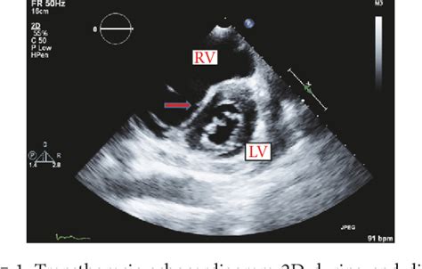
lv d shape | d shaped left ventricle function lv d shape In normal patients, the eccentricity index is 1.0 at both end-diastole and end-systole. Just remember that the left ventricle should look circular . See more Izsaki savas domas. Uzzini kur skatīt jaunākās filmas un seriālus online latviski vai ar subtitriem latviešu valodā!
0 · d shaped left ventricular septum
1 · d shaped left ventricular dysfunction
2 · d shaped left ventricle function
1. What is Red Mage's Playstyle? Red Mage is a magical DPS that primarily does damage by casting spells to build up its Black and White Mana gauges to 50|50, and then spends that mana to use an enchanted melee combo with powerful finisher spells.
To start looking for the D Sign on echo, you will need to obtain a Parasternal Short-Axis View of the heart. Click HEREfor the video series on how to get the cardiac views of the heart on echocardiography. See moreThe Eccentricity Index (EI) is an echocardiographic measurement of the left ventricle that can quantify the amount of right ventricular strain and overload affecting the left ventricle. It is described by Ryan et aland is calculated by taking two measurements . See moreIn patients with right ventricular Pressure overload, there is a significant elevation of pressure in the right ventricle throughout systole AND diastole. Because of this, the right ventricle will push in on the septum in both systole and diastole causing the left ventricle to be D . See moreIn normal patients, the eccentricity index is 1.0 at both end-diastole and end-systole. Just remember that the left ventricle should look circular . See more

In right ventricular Volumeoverload, only the diastolic phase is affected and the systolic phase is spared. The thought is that, in right . See more Together, these transgastric midpapillary short-axis images capture the classic echocardiographic finding of a “D”-shaped left ventricle (LV) . Flattening of the interventricular septum detected during echocardiographic examination is called D-shaped left ventricle. We present a case of an elderly male of African .The “D Sign” is an ultrasound/echo finding that shows the left ventricle as a D-shaped structure. It is a result of right ventricular overload causing a shift of the septum towards the left side of the heart. The “D-sign” can be the result of either right ventricular Pressure and/or Volume overload.
Together, these transgastric midpapillary short-axis images capture the classic echocardiographic finding of a “D”-shaped left ventricle (LV) secondary to septal flattening in the setting of right ventricular dysfunction. Flattening of the interventricular septum detected during echocardiographic examination is called D-shaped left ventricle. We present a case of an elderly male of African descent, who presented with increased shortness of breath.D-shaped left ventricle (D-LV), is an interesting echocardiographic finding in PH and is the result of structural distortion of the interventricular septum caused by an abnormal pressure gradient between the left and right ventricles. (D) Pericardial pressure increases more steeply with left-heart filling at higher right heart volumes, demonstrating how RV-LV interdependence alters apparent chamber stiffness.
d shaped left ventricular septum
The right ventricle—structural and functional importance for anaesthesia and intensive care. E Murphy 1, B Shelley 1,∗. Author information. Article notes. Copyright and License information. PMCID: PMC7808065 PMID: 33456839. Learning objectives. By .
Left ventricular (LV) diastolic function is characterized by LV relaxation, chamber stiffness, and early diastolic recoil, all of which determine LV filling pressure. Echocardiographic signals significantly associated with LV relaxation are mitral annulus early diastolic velocity (e′), LV strain rate during isovolumic relaxation (SR IVR .The left ventricular (LV) eccentricity index is calculated as the ratio of the 2 minor LV axes: LV lateral dimension (A) over the anterior-posterior (B) dimension. This index in systole or diastole reflects compression of the LV by the pressure- or volume-loaded RV, respectively.
Abstract. Background. Little is known about the impact of diastolic interventricular septal flattening on the clinical outcome in patients with severe tricuspid regurgitation. This study sought to evaluate the association of diastolic interventricular septal flattening with clinical outcome in patients with severe tricuspid regurgitation. Among them, D-shaped LV is one of echocardiographic parameters suggesting the presence of elevated RV pressure. An abnormal pressure gradient between LV and RV can lead to D-shaped LV. This can be calculated using the eccentricity index and is primarily used to separate patients with RV pressure from those with volume overload.The “D Sign” is an ultrasound/echo finding that shows the left ventricle as a D-shaped structure. It is a result of right ventricular overload causing a shift of the septum towards the left side of the heart. The “D-sign” can be the result of either right ventricular Pressure and/or Volume overload.Together, these transgastric midpapillary short-axis images capture the classic echocardiographic finding of a “D”-shaped left ventricle (LV) secondary to septal flattening in the setting of right ventricular dysfunction.
d shaped left ventricular dysfunction
Flattening of the interventricular septum detected during echocardiographic examination is called D-shaped left ventricle. We present a case of an elderly male of African descent, who presented with increased shortness of breath.
D-shaped left ventricle (D-LV), is an interesting echocardiographic finding in PH and is the result of structural distortion of the interventricular septum caused by an abnormal pressure gradient between the left and right ventricles. (D) Pericardial pressure increases more steeply with left-heart filling at higher right heart volumes, demonstrating how RV-LV interdependence alters apparent chamber stiffness.
nike handschoenen jd
The right ventricle—structural and functional importance for anaesthesia and intensive care. E Murphy 1, B Shelley 1,∗. Author information. Article notes. Copyright and License information. PMCID: PMC7808065 PMID: 33456839. Learning objectives. By .Left ventricular (LV) diastolic function is characterized by LV relaxation, chamber stiffness, and early diastolic recoil, all of which determine LV filling pressure. Echocardiographic signals significantly associated with LV relaxation are mitral annulus early diastolic velocity (e′), LV strain rate during isovolumic relaxation (SR IVR .
The left ventricular (LV) eccentricity index is calculated as the ratio of the 2 minor LV axes: LV lateral dimension (A) over the anterior-posterior (B) dimension. This index in systole or diastole reflects compression of the LV by the pressure- or volume-loaded RV, respectively. Abstract. Background. Little is known about the impact of diastolic interventricular septal flattening on the clinical outcome in patients with severe tricuspid regurgitation. This study sought to evaluate the association of diastolic interventricular septal flattening with clinical outcome in patients with severe tricuspid regurgitation.
d shaped left ventricle function

FileBase.lv. FileBase.lv bija viens no lielākājiem pēc reģistrēto lietotāju skaita Latvijas krievvalodīgo auditorijas portāls, kurš pakāpeniski audzēja savu auditoriju. Šis resurss sniedz lielu daudzumu filmu krievu valodā, kā arī daudzus citus krievu torrentus. BitHack.lv
lv d shape|d shaped left ventricle function


